Research

Research

The ubiquitylation system

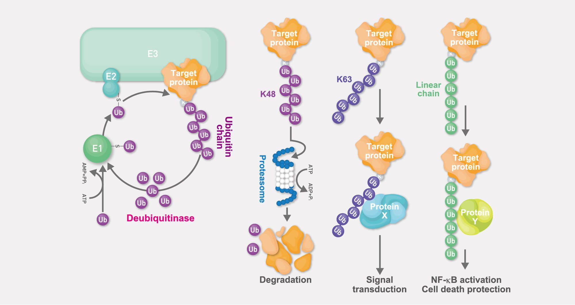
Ubiquitylation is one of the major post-translational modification systems, in which, via a cascade of reactions catalyzed by three types of enzymes (termed “E1”, “E2”, and “E3” enzymes), the E3 (ubiquitin ligase) target-specifically conjugates ubiquitin to substrate proteins.

Although originally the ubiquitin system had been identified as part of the cellular system of energy-dependent proteolysis (“ubiquitin-proteasome system”), the ubiquitin system is now well recognized as one of the most sophisticated reversible post-translational modification systems that regulates protein function in a wide variety of ways.

When compared to other post-translational modifications, the ubiquitin system shows several unique characteristics. They are:

  1. Ubiquitin is a proteineous post-translational modifier. This stands in stark contrast to most other post-translational modifiers which are small functional groups such as a phosphate, methyl, or acetyl groups.
  2. In most cases, a target protein’s function is modulated by modification with ubiquitin chains, i.e., polymeric forms of ubiquitin.
  3. Various types of ubiquitin chains exist in cells, and the type of the ubiquitin chain determines the functional mode of protein regulation; for this reason, ubiquitin pathways can exert a wide variety of functions.

For a long time it had been believed that ubiquitin chains are generated exclusively via one of ubiquitin’s seven lysine (Lys-linked) residues. In spite of this reigning paradigm, we discovered a brand-new type of ubiquitin chain: the linear ubiquitin chain. It is linked not via any of the Lys residues, but via the N-terminal methionine residue (Met1-linked). In addition to promoting pathophysiological analyses of these linear ubiquitin chains, we also discovered that abnormal generation of linear chains underlies various diseases including cancer and inflammatory diseases.

Thumbnail
1

Functional analysis of linear ubiquitin chains

Thumbnail
2

Role of linear ubiquitin chains in chronic inflammation and cancer

Thumbnail
3

Screening to identify drugs that control the function of LUBAC

Thumbnail
4

Analysis of intracellular iron dynamics and ferroptosis

1
Functional analysis of linear ubiquitin chains

Thumbnail

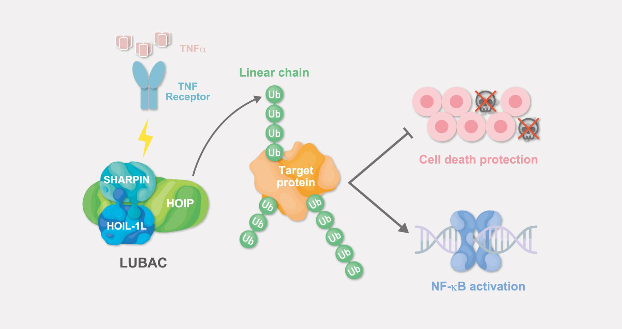
So far, the LUBAC ubiquitin ligase complex is the only known (E3) ubiquitin ligase that generates linear ubiquitin chains.

Linear ubiquitin chains generated by LUBAC are involved in activation of the transcription factor NF-κB induced by various extracellular stimuli such as TNF-α, and also play a crucial role in suppression of programmed cell death.

By elucidating mechanisms underlying the regulation of LUBAC and identifying new proteins targets of LUBAC, we are currently exploring yet unidentified roles of linear ubiquitin chains and their contribution to the onset of disease.

Publications

LUBAC accelerates B-cell lymphomagenesis by conferring B cells resistance to genotoxic stress.

Jo, T., Nishikori, M., Kogure, Y., Arima, H., Sasaki, K., Sasaki, Y., Nakagawa, T., Iwai, F., Momose, S., Shiraishi, A., Kiyonari, H., Kagaya, N., Onuki, T., Shin-ya, K., Yoshida, M., Kataoka, K., Ogawa, S., Iwai, K. and Takaori-Kondo, A.

Blood 136(6): 684-697, 2020. DOI

The HOIL-1L ligase modulates immune signaling and cell death via mono-ubiquitination of LUBAC

Fuseya, Y., Fujita, H., Kim, M., Ohtake, F., Nishide, A., Sasaki, K., Saeki, Y., Tanaka, K., Takahashi, R. and Iwai, K.

Nature Cell Biology 22(6): 663-673, 2020. DOI

Modulation of autoimmune pathogenesis by T cell-triggered inflammatory cell death.

Sasaki, K., Himeno, A., Nakagawa, T., Sasaki, Y., Kiyonari, H. and Iwai, K.

Nature Commun. 10(1):3878, 2019. DOI

Cooperative domain formation by homologous motifs in HOIL-1L and SHARPIN plays crucial roles in LUBAC stabilization.

Fujita, H., Tokunaga, A., Shimizu, S., Whiting, A. L., Aguilar-Alonso, F., Takagi, K., Walinda, E., Sasaki, Y., Shimokawa, T., Mizushima, T., Ohki, I., Ariyoshi, M., Tochio, H., Bernal, F., Shirakawa, M., and Iwai, K.

Cell Reports 23(4):1192-1204, 2018. DOI

Crucial role of LUBAC-mediated inhibition of programmed cell death in TLR4-mediated B cell responses and B1b cell development.

Sasaki, Y. and Iwai, K.

J. Immunol. 200(10):3438-3449, 2018. DOI

Differential involvement of the NZF domains of SHARPIN and HOIL-1L in LUBAC-mediated cell death protection.

Shimizu, S., Fujita, H., Sasaki, Y., Tsuruyama, T., Fukuda, K., and Iwai, K.

Mol. Cell. Biol. 36:1569-1583, 2016. DOI

IFN-gamma or IFN-alpha ameliorates chronic proliferative dermatitis by inducing expression of linear ubiquitin chain assembly complex.

Tamiya, H., Terao, M., Takiuchi, T., Nakahara, M., Sasaki, Y., Katayama, I., Yoshikawa, H., and Iwai K.

J. Immunol. 192:3793-3804, 2014.

Mechanism underlying IKK activation mediated by the linear ubiquitin chain assembly complex (LUBAC).

Fujita, H., Rahighi, S., Akita, M., Kato, R., Sasaki, Y., Wakatsuki, S., and Iwai, K.

Mol. Cell. Biol. 34:1322-1335, 2014.

Suppression of LUBAC-mediated linear ubiquitination by a specific interaction between LUBAC and the deubiquitinases CYLD and OTULIN.

Takiuchi, T., Nakagawa, T., Tamiya, H., Fujita, H., Sasaki, Y., Saeki, Y., Takeda, H., Sawasaki, T., Buchberger, A., Kimura, T., and Iwai. K.

Genes Cells. 19:254-272, 2014.

Defective immune responses in mice lacking LUBAC-mediated linear ubiquitination in B cells.

Sasaki, Y., Sano, S., Nakahara, M., Murata, S., Kometani, K., Aiba, Y., Sakamoto, S., Watanabe, Y., Tanaka, K., Kurosaki, K. and Iwai, K.

EMBO J. 32: 2463- 2476, 2013.

Specific Recognition of Linear Ubiquitin Chains by the HOIL-1L NZF domain.

Sato, Y., Fujita, H., Yoshikawa, A.,Yamashita, M., Yamagata, A., Kaiser, S. E., Iwai, K., and Fukai, S.

Proc. Natl. Acad. Sci. USA 108:20520-20525, 2011.

SHARPIN is a component of the NF-κB activating linear ubiquitin chain assembly complex.

Tokunaga, F., Nakagawa, T., Nakahara, M., Saeki, Y., Taniguchi, M., Sataka, S.-I., Tanaka, K., Nakano, H., and Iwai, K.

Nature 471:633-636, 2011.

Involvement of linear polyubiquitination of NEMO in NF-κB activation.

Tokunaga, F., Sakata, S.-I., Saeki, Y., Satomi, Y., Kirisako, T., Kamei, K., Nakagawa, T., Kato, M., Murata, S., Yamaoka, S., Yamamoto, M., Akira, S., Takao, T., Tanaka, K. and Iwai, K.

Nature Cell Biology 11:123-132, 2009.

A ubiquitin ligase complex assembles linear polyubiquitin chains.

Kirisako, T., Kamei, K., Murata, S., Kato, M., Fukumoto, H., Kanie, K., Sano, S. Tokunaga, F., Tanaka, K. and Iwai, K.

EMBO J. 25: 4877–4887, 2006.

Identification of the ubiquitin-protein ligase that recognizes oxidized IRP2.

Yamanaka, K., Ishikawa, H., Megumi, Y., Tokunaga, F., Kanie, M., Rouault, T.A., Morishima, I., Minato, N., Ishimori, K. and Iwai, K.

Nature Cell Biology 5:336-340, 2003.

View all

2
Role of linear ubiquitin chains in chronic inflammation and cancer

Thumbnail

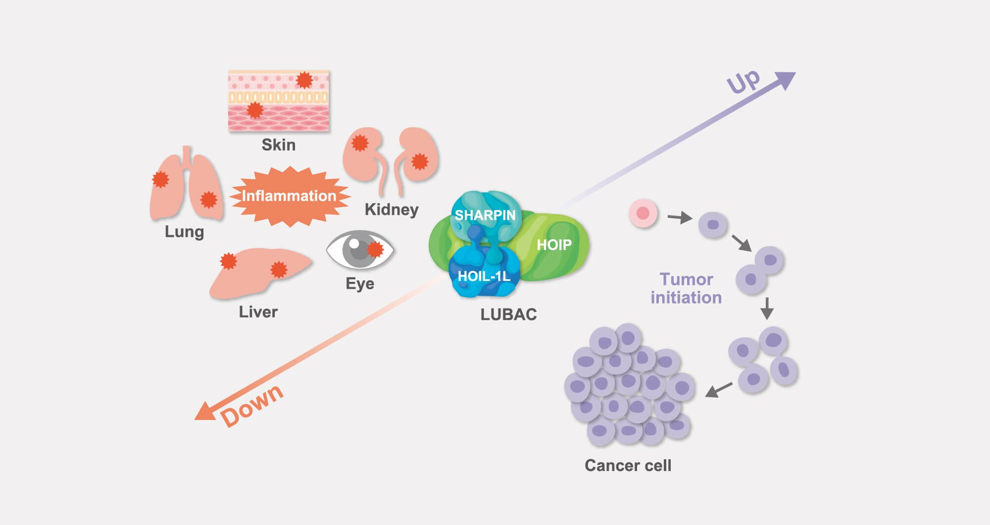
We have shown that, by regulating intracellular signal transduction systems, linear ubiquitylation plays crucial roles in both onset and maintenance (and even exacerbation) of the inflammatory environment in tissues in many immune and inflammatory diseases including cancer.

By utilizing organ-specific LUBAC-hyperactive and -deficient models, we are currently aiming at clarifying even more detailed physiological functions of the linear ubiquitin chain at the tissue level and at the level of individual animals. At the same time, we are analyzing new mechanisms that act in the pathogenesis of inflammatory diseases by applying various experimental approaches.

Publications

LUBAC accelerates B-cell lymphomagenesis by conferring B cells resistance to genotoxic stress.

Jo, T., Nishikori, M., Kogure, Y., Arima, H., Sasaki, K., Sasaki, Y., Nakagawa, T., Iwai, F., Momose, S., Shiraishi, A., Kiyonari, H., Kagaya, N., Onuki, T., Shin-ya, K., Yoshida, M., Kataoka, K., Ogawa, S., Iwai, K. and Takaori-Kondo, A.

Blood 136(6): 684-697, 2020. DOI

Modulation of autoimmune pathogenesis by T cell-triggered inflammatory cell death.

Sasaki, K., Himeno, A., Nakagawa, T., Sasaki, Y., Kiyonari, H. and Iwai, K.

Nature Commun. 10(1):3878, 2019. DOI

IFN-gamma or IFN-alpha ameliorates chronic proliferative dermatitis by inducing expression of linear ubiquitin chain assembly complex.

Tamiya, H., Terao, M., Takiuchi, T., Nakahara, M., Sasaki, Y., Katayama, I., Yoshikawa, H., and Iwai K.

J. Immunol. 192:3793-3804, 2014.

SHARPIN is a component of the NF-κB activating linear ubiquitin chain assembly complex.

Tokunaga, F., Nakagawa, T., Nakahara, M., Saeki, Y., Taniguchi, M., Sataka, S.-I., Tanaka, K., Nakano, H., and Iwai, K.

Nature 471:633-636, 2011.

SHARPIN forms a linear ubiquitin ligase complex regulating NF-κB activity and apoptosis.

Ikeda, F., Deribe, Y. L., Skånland, S.S., Stieglitz, B., Grabbe, C., Franz-Wachtel, M., van Wijk, S.J.L., Goswami, P., Nagy, V., Terzic, J., Tokunaga, F., Androulidaki, A., Nakagawa, T., Pasparakis, M., Iwai, K., Sundberg, J.P., Rittinger, K., Schaefer, L., Macek, B. and Dikic, I.

Nature 471:637-641, 2011.

View all

3
Screening to identify drugs that control the function of LUBAC

Thumbnail

In numerous previous studies, we and other groups have clearly demonstrated that both augmented and attenuated function of LUBAC can provoke various diseases.

We therefore aim at developing new therapeutics targeting LUBAC. Drugs that have the potency to inhibit LUBAC activity are likely to be suitable as anticancer drugs, whereas drugs that increase LUBAC activity are thought to be promising antibacterial drugs.

Based on our findings, we are now actively conducting drug screening assays to develop both LUBAC inhibitors and activators.

Publications

LUBAC accelerates B-cell lymphomagenesis by conferring B cells resistance to genotoxic stress.

Jo, T., Nishikori, M., Kogure, Y., Arima, H., Sasaki, K., Sasaki, Y., Nakagawa, T., Iwai, F., Momose, S., Shiraishi, A., Kiyonari, H., Kagaya, N., Onuki, T., Shin-ya, K., Yoshida, M., Kataoka, K., Ogawa, S., Iwai, K. and Takaori-Kondo, A.

Blood 136(6): 684-697, 2020. DOI

Cooperative domain formation by homologous motifs in HOIL-1L and SHARPIN plays crucial roles in LUBAC stabilization.

Fujita, H., Tokunaga, A., Shimizu, S., Whiting, A. L., Aguilar-Alonso, F., Takagi, K., Walinda, E., Sasaki, Y., Shimokawa, T., Mizushima, T., Ohki, I., Ariyoshi, M., Tochio, H., Bernal, F., Shirakawa, M., and Iwai, K.

Cell Reports 23(4):1192-1204, 2018. DOI

Gliotoxin Suppresses NF-κB Activation by Selectively Inhibiting Linear Ubiquitin Chain Assembly Complex (LUBAC).

Sakamoto, H., Egashira, S., Saito, N., Kirisako, T., Miller, S., Sasaki, Y., Matsumoto, T., Shimonishi, M., Komatsu, T., Terai, T., Ueno, T., Hanaoka, K., Kojima, H., Okabe, T., Wakatsuki, S., Iwai, K., (coreesonding author) and Nagano, T.

ACS Chem Biol. 10:675-681, 2015.

Essential role of the linear ubiquitin chain assembly complex in lymphoma revealed by rare germline polymorphisms.

Yang, Y., Schmitz, R., Mitala, J. J. Jr., Whiting, A., Xiao, W., Ceribelli, M., Wright, G. W., Zhao, H., Yang, Y., Xu, W., Rosenwald, A., Ott, G., Gascoyne, R. D., Connors, J. M., Rimsza, L. M., Campo, E., Jaffe, E. S., Delabie, J., Smeland, E. B., Braziel, R. M., Tubbs, R. R., Cook, J. R., Weisenburger, D. D., Chan, W. C., Wiestner, A., Kruhlak, M. J., Iwai, K., Bernal, F., and Staudt, L. M.

Cancer Discovery 4:480-493, 2014.

View all

4
Analysis of intracellular iron dynamics and ferroptosis

Thumbnail

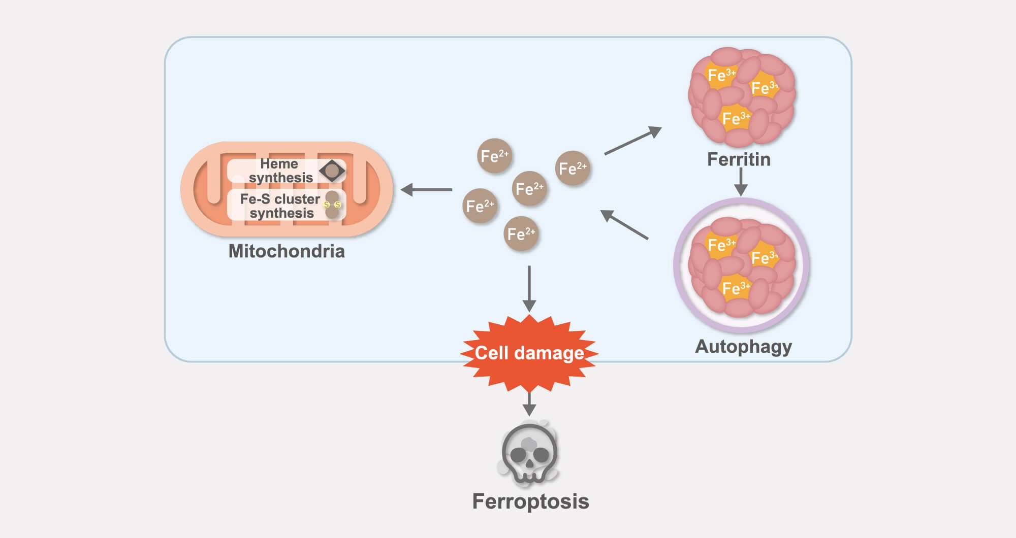
Iron is an essential nutrient for almost all living organisms. Conversely, it can also be toxic when present in excess. For this reason, intracellular iron metabolism must be tightly regulated.

We are studying the mechanism of iron homeostasis through the analysis of mitochondria, in which iron is integrated into iron-binding prosthetic groups such as the heme group, iron-sulfur clusters, and the iron-storage protein ferritin.

In addition, we are – from an iron-based perspective – extensively dissecting the molecular mechanisms underlying ferroptosis, which is an iron-dependent form of cell death that has been attracting tremendous attention in recent years.

Publications

Possible involvement of iron-induced oxidative insults in neurodegeneration.

Asano, T., Koike, M., akata, S.-I., Takeda, Y., Nakagawa, T., Hatano, T., Ohashi, S., Funayama, M., Yoshimi, K., Asanuma, M., Toyokuni, S., Mochizuki, H., Uchiyama, Y., Hattori, N., and Iwai, K.

Neurosci. Lett. 588:29-35, 2015.

Iron-Induced Dissociation of the Aft1p Transcriptional Regulator from Target Gene Promoters is an Initial Event in Iron-Dependent Gene Suppression.

Ueta, R., Fujiwara, N., Iwai, K. (Correspondence author) and Yamaguchi-Iwai, Y.

Mol. Cell. Biol. 32:4998-5008, 2012.

Distinct mechanisms of ferritin delivery to lysosomes in iron-depleted and iron-replete cells.

Asano, T., Komatsu, M., Yamaguchi-Iwai, Y., Ishikawa, F., Mizushima, N. and Iwai, K.

Mol. Cell. Biol. 31:2040-2052, 2011.

The FBXL5-IRP2 axis is integral to control of iron metabolism in vivo.

Moroishi, T., Nishiyama, M., Takeda, Y., Iwai, K. and Nakayama, K.I.

Cell Metabolism 14: 339–351, 2011.

Mechanism underlying the iron-dependent nuclear export of the iron-responsive transcription factor Aft1p in Saccharomyces cerevisiae.

Ueta, R., Fujiwara, N., Iwai, K. (corresponding author) and Yamaguchi-Iwai, Y.

Mol. Biol. Cell 18:2980-2990, 2007.

Involvement of heme regulatory motif in heme-mediated ubiquitination and degradation of IRP2.

Ishikawa, H., Kato, M., Hori, H., Ishimori, K., Kirisako, T., Tokunaga, F. and Iwai, K.

Molecular Cell 19:171-181, 2005.

Iron-dependent oxidation, ubiquitination, and degradation of iron regulatory protein 2: Implications for degradation of oxidized proteins.

Iwai, K., Drake, S. K., Wehr, N. B., Weissman, A. M., LaVaute, T. M., Minato, N., Klausner, R.D., Levine, R.L. and Rouault, T.A.

Proc. Natl. Acad. Sci. USA 95:4924-4928, 1998.

Requirements for iron-regulated degradation of the RNA binding protein, iron regulatory protein 2.

Iwai, K., Klausner, R.D. and Rouault, T.A.

EMBO. J. 14:5350-5357, 1995.

View all